While the link between a high-fat diet and memory impairment has been well documented, the exact reason for this association has remained elusive. Now, a new study by researchers from The Barrientos Laboratory at Ohio State University has not only uncovered ways in which fatty foods affect brain cells, it has also identified one possible means of protecting those cells from this damage.
The Study
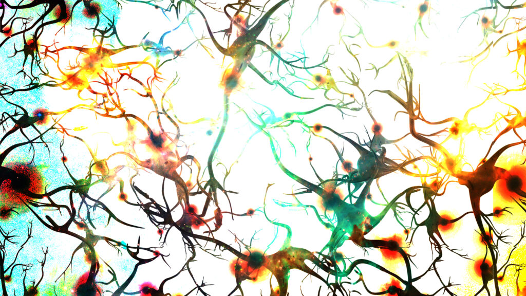
The researchers focused on microglia, cells in the brain that promote inflammation, and hippocampal neurons, which are important for learning and memory. They exposed microglia and neurons from rats to palmitic acid, the most abundant saturated fatty acid found in high-fat foods like lard, shortening, meat, and dairy products. They found that palmitic acid prompted gene expression changes linked to an increase in inflammation in both microglia and neurons, though microglia had a wider range of affected inflammatory genes.
Perhaps even more importantly, the researchers found that pre-treatment of these cells with a dose of DHA, one of two omega-3 fatty acids found in seafood and available in supplement form, had a strong protective effect against the increased inflammation in both cell types.
“Previous work has shown that DHA is protective in the brain and that palmitic acid has been detrimental to brain cells, but this is the first time we’ve looked at how DHA can directly protect against the effects of palmitic acid in those microglia, and we see that there is a strong protective effect,” said Michael Butler, PhD, first author of the study.
In another set of experiments, the researchers looked at how a diet high in saturated fat influenced signaling in the brains of aged mice by observing another microglial function called synaptic pruning. Microglia monitor signal transmission among neurons and nibble away excess synaptic spines, the connection sites between axons and dendrites, to keep communication at an ideal level.
Microglia were exposed to mouse brain tissue from animals that had been fed either a high-fat diet or regular chow for three days. The microglia ate the synapses from the mice on the high-fat diet at a faster rate, suggesting the high-fat diet does something to those synapses that gives the microglia more reason to eat them.
“When we talk about the pruning…it’s like Goldilocks: It needs to be optimal – not too much and not too little,” said senior study author Ruth Barrientos, PhD. “With these microglia eating away too much too soon, it outpaces the ability for these spines to regrow and create new connections, so memories don’t solidify or become stable.”
Conclusions
“The cool thing about this paper is that for the first time, we’re really starting to tease these things apart by cell type,” said Barrientos. “Our lab and others have often looked at the whole tissue of the hippocampus to observe the brain’s memory-related response to a high-fat diet. But we’ve been curious about which cell types are more or less affected by these saturated fatty acids, and this is our first foray into determining that.”
From here, the researchers plan to conduct further experiments to expand on their findings related to synaptic pruning and mitochondria function and to see how the palmitic acid and DHA effects play out in primary brain cells from young versus aged animals.


