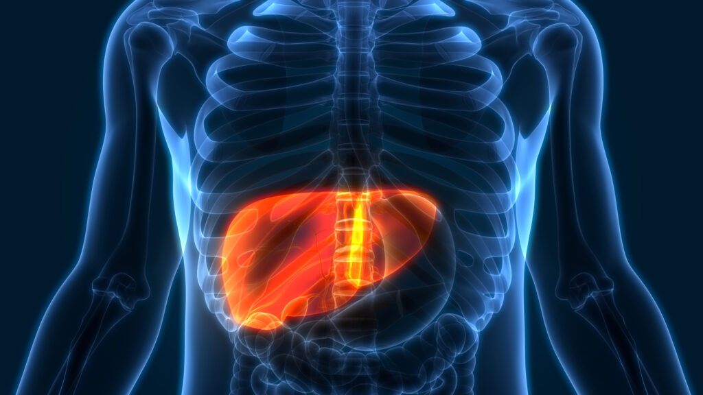
Patients with non-alcoholic fatty liver disease (NAFLD) have increased odds of depression, dementia, and other neurological conditions, according to a study in the Journal of Hepatology.
NAFLD is usually caused by a high-sugar, high-fat diet, which has been linked to brain fog, mood imbalances, and other brain-related symptoms on its own. Now, there is a direct link between NAFLD — an increasingly common liver disease affecting 25 percent of the population — and suboptimal brain function.
A buildup of fat in the liver is associated with a decrease in oxygen in the brain, as well as brain tissue inflammation, said the researchers. Both conditions are present in those with serious brain disorders.
Study Details
For the study, mice were divided into two groups and given either a low-fat diet (10 percent fat or less) or a high-fat diet (55 percent fat). The latter group’s food was designed to mimic a diet high in processed foods and sugary drinks.
At the end of 16 weeks, mice on the higher-fat diet became obese, and they developed NAFLD, along with brain dysfunction and insulin resistance. The mice in the low-fat group did not have any of the same problems — no NAFLD, brain problems, or insulin resistance.
“It is very concerning to see the effect that fat accumulation in the liver can have on the brain, especially because it often starts off mild and can exist silently for many years without people knowing they have it,” said lead author Dr. Anna Hadjihambi, sub-team lead in the Liver-Brain Axis group at the Roger Williams Institute of Hepatology and honorary lecturer at King’s College London.
The Role of MCT1
Researchers wanted to know if Monocarboxylate Transporter 1 (MCT1), a protein involved in energy substrate transport, played a part in the liver- and brain-damaging effects of high-fat, high-sugar diets. So, they bred mice to have reduced levels of MCT1 and discovered the mice had no fat accumulation and zero signs of brain degeneration — despite being fed the same unhealthy 55 percent-fat diet. MCT1 appears to be a key element in the liver-brain axis and may help scientists develop therapies to target NAFLD.
Conclusion
“In conclusion, this study provides evidence indicating a key role of NAFLD in inducing low-grade brain tissue hypoxia and inflammation, as well as cerebrovascular, glial, metabolic, and behavioral alterations. Such effects are expected to persist chronically or even worsen with disease progression, leading to the early stages of NAFLD-induced brain dysfunction, while increasing the risk of neurodegenerative conditions, such as Alzheimer’s disease, that share the above pathophysiological mechanisms.42 Mct1 haploinsufficient mice, despite fat accumulation in adipose tissue, were protected from NAFLD and the above detrimental cerebral alterations, emphasizing the importance of the liver–brain axis in developing cognitive decline observed in obese individuals. Finally, this protective phenotype indicates the potential of MCT1 as a novel therapeutic target for preventing and/or treating NAFLD and the associated multifactorial encephalopathy.”


