Data shows a relationship between environmental toxicants and diabetes – so, what can you do to protect your patients?
The first Global Report on Diabetes, recently published by the World Health Organization (WHO),[1] highlighted several alarming statistics: the incidence of diabetes has doubled since 1980, and as of 2012, it was the eighth leading cause of death worldwide among both sexes, and the fifth leading cause of death in women. Diabetes was responsible for 1.5 million deaths in 2012, more than 43% of which occurred in individuals under 70 years of age. And the financial impact of diabetes is even more shocking – the cost of this disease in the U.S. increased from $116 billion in 2007 to $237 billion in 2017, and per person, costs an average of $16,752 yearly.[2]
As the report explains, the increase in type-2 diabetes (T2DM) mirrors the likewise alarming increasing prevalence of obesity, with one in three adults over the age of 18 overweight and one in ten being obese. Excessive body fat is the strongest risk factor for the development of T2DM, while other risk factors include higher waist circumference, higher body mass index (BMI), and being an active smoker. Dietary patterns that include a high intake of saturated and total fat, low consumption of fiber, and high intake of sugar-sweetened beverages are also risk factors for T2DM and/or excess body weight. Early childhood nutrition also affects the risk of development of T2DM later in life.
One factor not considered in the WHO overview of diabetes, however, is the potential impact of environmental risk factors on the development of the disease.
Environmental toxicants and diabetes
Exposure to environmental toxicants can occur through the air, contact with the skin, contaminated drinking water, and food. Many environmental pollutants do not degrade over time, and thus continue to accumulate in the environment; these chemicals are classified as persistent organic pollutants (POPs). Similarly, they are persistent in the human body, as their lipophilic nature draws them into fatty tissue like our subcutaneous fat as well as the brain,[3],[4] from which they are slowly released into the bloodstream even if we are not exposed to them on an ongoing basis.
In a survey of 2,016 adult participants, performed by the National Health and Nutrition Examination Survey (NHANES), diabetes prevalence was strongly associated with blood concentrations of six POPs.[5] Interestingly, obese persons who did not have elevated POPs were not at elevated risk of diabetes. This suggests that the POP exposure, rather than the obesity, was responsible for the increased risk.[6]
Environmental toxicants have the ability to affect genetic transcription, disrupting DNA methylation and altering the organization of chromatin which normally serves to prevent DNA damage and control gene expression.[7] Exposure to environmental pollutants can lead to epigenetic changes, which have the potential to affect more than just one generation.[8],[9] In addition to obesity and diabetes, environmental toxicants have been associated with several types of cancer,[10] respiratory disease,[11] cardiovascular disease,[12] infertility,[13] allergies,[14] autoimmune disease,[15] and many other conditions.[16]
Endocrine-disrupting chemicals (EDCs) wreak havoc on the hormones, and therefore can have an adverse effect on male and female reproductive health, sexual development, thyroid function, metabolism, and increase risk of breast and prostate cancers.[17] EDCs include pollutants like organochlorinated pesticides and industrial chemicals, plastics and plasticizers, fuels, and many other chemicals.[18]
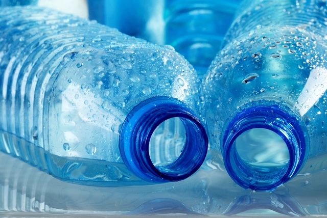
Exposure to bisphenol A (BPA), a known EDC, has been linked to obesity, diabetes, cardiovascular diseases, polycystic ovarian disease (PCOS), and low sperm count.[19],[20] BPA is found in the polycarbonate plastics that are used in many types of food and drink packaging including food storage containers, water bottles, baby bottles, and the internal protective coating of cans that hold food.[21] The degree to which BPA leaches from these materials into food depends mostly on the temperature it is exposed to, with hotter food leading to higher amounts. Non-food sources of BPA also are significant with thermal papers and dental materials being sources of exposure as well.[22],[23] BPA can also be found in the breast milk of mothers with high levels of exposure, and readily transfers to the infant.[24]
BPA inhibits adipocytes from releasing adiponectin, an adipokine that is a key regulator of insulin sensitivity[25] and helps to protect humans from obesity, diabetes, and heart disease.[26] BPA has been shown to affect the function of both the insulin-releasing β-cells and the glucagon releasing α-cells in the pancreas.[27] Animal studies have shown that exposure to BPA in utero may contribute to the development of diabetes in the offspring and aggravates insulin resistance associated with pregnancy in the mother.[28] In data analyzing 4,389 adults with diabetes, higher urinary BPA levels were associated with higher hemoglobin A1c (HbA1c) levels.[29] In another recent study, increased exposure to BPA was associated with insulin resistance in overweight or obese children.[30]
Dioxins are POPs that also are classified as EDCs. Dioxins are also stored in adipose tissue and accumulate in the fat of the animals we eat, as well as our bodies.[31] More than 90% of human dioxin exposure is through food, with roughly 50% being from meat and dairy.[32] Much like BPA, dioxins also transfer to infants in utero and via the breast milk.[33] Dioxins have been shown to reduce expression of glucose transporters, decreasing glucose uptake by adipose tissue, the liver and the pancreas.[34] They contribute to mitochondrial dysfunction, which in pancreatic cells can adversely affect the release of insulin.[35] In both men and women, a high serum dioxin level was shown to be an independent risk factor for the development of diabetes, independent of age and weight.[36] Other studies support these findings, however there are some inconsistencies which suggest other factors also may be at play.[37],[38]
In addition to BPA and dioxins, environmental chemicals such as polycyclic aromatic hydrocarbons and volatile organic compounds also cause oxidative damage in the body.[39] Oxidative stress contributes to insulin resistance and β-cell dysfunction[40] as well as the complications associated with diabetes.[41]
Natural agents that minimize harm
Needless to say, lifestyle changes to reduce exposure to harmful compounds such as these may have beneficial effects on many aspects of health as well as the risk of diabetes. However, certain natural products also have data that suggests they may help mitigate the harm caused by environmental toxicants.
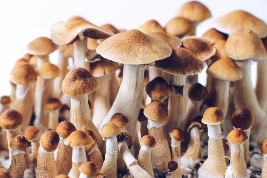
Chlorella, well known for its detoxifying effects, has been shown to reduce levels of dioxin in human breast milk, simultaneously increasing levels of the protective immuglobulins which help reduce the risk of infant infection.[42] In humans, intake of chlorella has been shown to affect the transcription of genes associated with fat metabolism and insulin signaling, simultaneously reducing body fat percentage, serum total cholesterol, and fasting blood glucose levels.[43] In patients with non-alcoholic fatty liver disease (NAFLD), also common amongst individuals with T2DM, chlorella in combination with vitamin E also significantly improved fasting blood sugar and lipid profiles.[44]
Oregon grape
Berberine, a compound found in botanicals such as Oregon grape, goldenseal, and barberry, has a multitude of actions that may help reduce damage from these environmental compounds as well as clinical evidence of its benefits for diabetics.[45] It serves as an antioxidant and anti-inflammatory agent,[46] reduces insulin resistance, promotes insulin secretion, inhibits gluconeogenesis in the liver, and stimulates glycolysis in peripheral tissue cells.[47],[48] Berberine also helps balance the gut microbiota and regulates cholesterol production.[49]
Lipoic acid is one antioxidant that helps protect against BPA and dioxin toxicity which also has an array of evidence for its use in diabetes.[50],[51] Studies show that lipoic acid improves glucose utilization, reduces HbA1c levels, supports blood vessel health, and mitigates the peripheral neuropathy complications of diabetes.[52],[53],[54]
Milk thistle, a well-known hepatoprotective herb commonly used as a part of detoxification protocols, also has evidences it helps protect against the oxidative damage induced by environmental contaminants.[55],[56] Milk thistle has evidence both in vitro and in vivo of hypoglycemic and hypolipidemic properties.[57] In patients with both alcoholic liver cirrhosis and diabetes, milk thistle was shown to significantly reduce fasting blood glucose and HbA1c levels, also decreasing insulin requirement by 20%.[58] Human studies have shown milk thistle has synergistic effects on blood sugar when combined with berberine.[59] The antioxidant effects of milk thistle and silymarin, the family of active compounds found in milk thistle, also have been shown to help prevent the development of diabetic nephropathy in animal studies.[60]
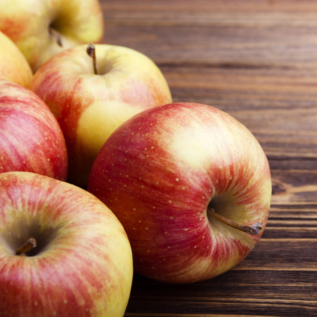
N-acetylcysteine (NAC), an antioxidant that also provides cysteine for the production of glutathione, has been found to attenuate the inflammation, oxidative damage, and cognitive dysfunction associated with BPA exposure.[61],[62] In animals, NAC also has been shown to reduce hyperglycemia and diabetes severity,[63] also protecting against hyperglycemia, hyperinsulinemia, and hepatic fat accumulation associated with high-fat diet feeding.[64] Quercetin, a natural compound found in foods such as apples, has evidence it helps mitigate oxidative damage to the liver and kidneys that can be caused by BPA.[65] Quercetin also has evidence that it helps prevent oxidative stress-related β-cell damage in animal models of diabetes.[66]
Summary
Environmental toxicants in our air, water, and food produce oxidative stress and contribute to insulin resistance, but natural substances can help mitigate the harm. Chlorella, berberine, lipoic acid, milk thistle, N-acetylcysteine, and quercetin are just a few potential substances which may exert protective effects in the face of exposure to environmental toxins and defend the body from related diabetes and metabolic dysfunction. Many of these agents can be used as supplements, and in addition to dietary and lifestyle changes, offer a holistic approach for protecting the body against chemical exposures.
Dr. Carrie Decker, ND graduated with honors from the National College of Natural Medicine (now the National University of Natural Medicine) in Portland, Oregon. Dr. Decker sees patients remotely, with a focus on gastrointestinal disease, mood imbalances, eating disorders, autoimmune disease, and chronic fatigue. Prior to becoming a naturopathic physician, Dr. Decker was an engineer, and obtained graduate degrees in biomedical and mechanical engineering from the University of Wisconsin-Madison and University of Illinois at Urbana-Champaign respectively. Dr. Decker continues to enjoy academic research and writing and uses these skills to support integrative medicine education as a writer and contributor to various resources. Dr. Decker supports Allergy Research Group as a member of their education and product development team.
Sources:
[1] World Health Organization. Global report on diabetes [Internet]. Geneva: World Health Organization; 2016 [cited 2018 Oct 8]. Available from: http://www.who.int/diabetes/global-report/en/
[2] Riddle MC, Herman WH. The Cost of Diabetes Care-An Elephant in the Room. Diabetes Care. 2018 May;41(5):929-932.
[3] La Merrill M, et al. Toxicological function of adipose tissue: focus on persistent organic pollutants. Environ Health Perspect. 2013 Feb;121(2):162-9.
[4] Dewailly E, et al. Concentration of organochlorines in human brain, liver, and adipose tissue autopsy samples from Greenland. Environ Health Perspect. 1999 Oct;107(10):823-8.
[5] Lee DH, et al. A strong dose-response relation between serum concentrations of persistent organic pollutants and diabetes: results from the National Health and Examination Survey 1999-2002. Diabetes Care. 2006 Jul;29(7):1638-44.
[6] Carpenter DO. Environmental contaminants as risk factors for developing diabetes. Rev Environ Health. 2008 Jan-Mar;23(1):59-74.
[7] Baccarelli A, Bollati V. Epigenetics and environmental chemicals. Curr Opin Pediatr. 2009 Apr;21(2):243-51.
[8] Feinberg AP, et al. Phenotypic plasticity and the epigenetics of human disease. Nature. 2007 May 24;447(7143):433-40.
[9] Skinner MK, et al. Epigenetic transgenerational actions of environmental factors in disease etiology. Trends Endocrinol Metab. 2010 Apr;21(4):214-22.
[10] Irigaray P, et al. Lifestyle-related factors and environmental agents causing cancer: an overview. Biomed Pharmacother. 2007 Dec;61(10):640-58.
[11] Khafaie MA, et al. Critical review of air pollution health effects with special concern on respiratory health. J Air Pollution Health. 2016 May 29;1(2):123-36.
[12] Bhatnagar A, et al. Environmental cardiology: studying mechanistic links between pollution and heart disease. Circ Res. 2006 Sep 29;99(7):692-705.
[13] Mendiola J, et al. Exposure to environmental toxins in males seeking infertility treatment: a case-controlled study. Reprod Biomed Online. 2008 Jun;16(6):842-50.
[14] Diaz-Sanchez D, et al. Diesel fumes and the rising prevalence of atopy: an urban legend? Curr Allergy Asthma Rep. 2003 Mar;3(2):146-52.
[15] Vojdani A, et al. A Potential Link between Environmental Triggers and Autoimmunity. Autoimmune Dis. 2014;2014:437231.
[16] Schug TT, et al. Endocrine disrupting chemicals and disease susceptibility. J Steroid Biochem Mol Biol. 2011 Nov;127(3-5):204-15.
[17] Schug TT, et al. Endocrine disrupting chemicals and disease susceptibility. J Steroid Biochem Mol Biol. 2011 Nov;127(3-5):204-15.
[18] Diamanti-Kandarakis E, et al. Endocrine-disrupting chemicals: an Endocrine Society scientific statement. Endocr Rev. 2009 Jun;30(4):293-342.
[19] Fenichel P, et al. Bisphenol A: an endocrine and metabolic disruptor. Ann Endocrinol (Paris). 2013 Jul;74(3):211-20.
[20] Ngwa EN, et al. Persistent organic pollutants as risk factors for type 2 diabetes. Diabetol Metab Syndr. 2015 Apr 30;7:41.
[21] Geens T, et al. A review of dietary and non-dietary exposure to bisphenol-A. Food Chem Toxicol. 2012 Oct;50(10):3725-40.
[22] Björnsdotter MK, et al. Bisphenol A and replacements in thermal paper: A review. Chemosphere. 2017 Sep;182:691-706.
[23] Fleisch AF, et al. Bisphenol A and related compounds in dental materials. Pediatrics. 2010 Oct;126(4):760-8.
[24] Mendonca K, et al. Bisphenol A concentrations in maternal breast milk and infant urine. Int Arch Occup Environ Health. 2014 Jan;87(1):13-20.
[25] Whitehead J, et al. Adiponectin–a key adipokine in the metabolic syndrome. Diabetes Obes Metab. 2006 May;8(3):264-80.
[26] Hugo ER, et al. Bisphenol A at environmentally relevant doses inhibits adiponectin release from human adipose tissue explants and adipocytes. Environ Health Perspect. 2008 Dec;116(12):1642-7.
[27] Ropero AB, et al. Bisphenol-A disruption of the endocrine pancreas and blood glucose homeostasis. Int J Androl. 2008 Apr;31(2):194-200.
[28] Alonso-Magdalena P, et al. Bisphenol A exposure during pregnancy disrupts glucose homeostasis in mothers and adult male offspring. Environ Health Perspect. 2010 Sep;118(9):1243-50.
[29] Silver MK, et al. Urinary bisphenol A and type-2 diabetes in U.S. adults: data from NHANES 2003-2008. PLoS One. 2011;6(10):e26868.
[30] Bertoli S, et al. Human bisphenol A exposure and the “diabesity phenotype.” Dose Response. 2015 Jul 31;13(3):1559325815599173.
[31] Schecter A, et al. Partitioning of dioxins, dibenzofurans, and coplanar PCBS in blood, milk, adipose tissue, placenta and cord blood from five American women. Chemosphere. 1998 Oct-Nov;37(9-12):1817-23.
[32] Charnley G, Doull J. Human exposure to dioxins from food, 1999-2002. Food Chem Toxicol. 2005 May;43(5):671-9.
[33] Todaka T, et al. Relationship between the concentrations of polychlorinated dibenzo-p-dioxins, polychlorinated dibenzofurans, and polychlorinated biphenyls in maternal blood and those in breast milk. Chemosphere. 2010 Jan;78(2):185-92.
[34] De Tata V. Association of dioxin and other persistent organic pollutants (POPs) with diabetes: epidemiological evidence and new mechanisms of beta cell dysfunction. Int J Mol Sci. 2014 May 5;15(5):7787-811.
[35] Maechler P, et al. In beta-cells, mitochondria integrate and generate metabolic signals controlling insulin secretion. Int J Biochem Cell Biol. 2006;38(5-6):696-709.
[36] Huang CY, et al. Association between dioxin and diabetes mellitus in an endemic area of exposure in Taiwan: a population-based study. Medicine (Baltimore). 2015 Oct;94(42):e1730.
[37] Steenland K, et al. Dioxin and diabetes mellitus: an analysis of the combined NIOSH and Ranch Hand data. Occup Environ Med. 2001 Oct;58(10):641-8.
[38] Calvert GM, et al. Evaluation of diabetes mellitus, serum glucose, and thyroid function among United States workers exposed to 2,3,7,8-tetrachlorodibenzo-p-dioxin. Occup Environ Med. 1999 Apr;56(4):270-6.
[39] Hong YC, et al. Community level exposure to chemicals and oxidative stress in adult population. Toxicol Lett. 2009 Jan 30;184(2):139-44.
[40] Evans JL, et al. Are oxidative stress-activated signaling pathways mediators of insulin resistance and beta-cell dysfunction? Diabetes. 2003 Jan;52(1):1-8.
[41] Ceriello A, Motz E. Is oxidative stress the pathogenic mechanism underlying insulin resistance, diabetes, and cardiovascular disease? The common soil hypothesis revisited. Arterioscler Thromb Vasc Biol. 2004 May;24(5):816-23.
[42] Nakano S, et al. Chlorella (Chlorella pyrenoidosa) supplementation decreases dioxin and increases immunoglobulin a concentrations in breast milk. J Med Food. 2007 Mar;10(1):134-42.
[43] Mizoguchi T, et al. Nutrigenomic studies of effects of Chlorella on subjects with high-risk factors for lifestyle-related disease. J Med Food. 2008 Sep;11(3):395-404.
[44] Ebrahimi-Mameghani M, et al. The Effect of Chlorella vulgaris Supplementation on Liver En-zymes, Serum Glucose and Lipid Profile in Patients with Non-Alcoholic Fatty Liver Disease. Health Promot Perspect. 2014 Jul 12;4(1):107-15.
[45] Lan J, et al. Meta-analysis of the effect and safety of berberine in the treatment of type 2 diabetes mellitus, hyperlipemia and hypertension. J Ethnopharmacol. 2015 Feb 23;161:69-81.
[46] Li Z, et al. Antioxidant and anti-inflammatory activities of berberine in the treatment of diabetes mellitus. Evid Based Complement Alternat Med. 2014;2014:289264.
[47] Wang Y, et al. Hypoglycemic and insulin-sensitizing effects of berberine in high-fat diet- and streptozotocin-induced diabetic rats. Metabolism. 2011 Feb;60(2):298-305.
[48] Pang B, et al. Application of berberine on treating type 2 diabetes mellitus. Int J Endocrinol. 2015;2015:905749.
[49] Han J, et al. Modulating gut microbiota as an anti-diabetic mechanism of berberine. Med Sci Monit. 2011 Jul;17(7):RA164-7.
[50] El-Beshbishy HA, et al. Lipoic acid mitigates bisphenol A-induced testicular mitochondrial toxicity in rats. Toxicol Ind Health. 2013 Nov;29(10):875-87.
[51] Koga T, et al. Restoration of dioxin-induced damage to fetal steroidogenesis and gonadotropin formation by maternal co-treatment with α-lipoic acid. PLoS One. 2012;7(7):e40322.
[52] Packer L, et al. Molecular aspects of lipoic acid in the prevention of diabetes complications. Nutrition. 2001 Oct;17(10):888-95.
[53] Poh ZX, Goh KP. A current update on the use of alpha lipoic acid in the management of type 2 diabetes mellitus. Endocr Metab Immune Disord Drug Targets. 2009 Dec;9(4):392-8.
[54] Sola S, et al. Irbesartan and lipoic acid improve endothelial function and reduce markers of inflammation in the metabolic syndrome: results of the Irbesartan and Lipoic Acid in Endothelial Dysfunction (ISLAND) study. Circulation. 2005 Jan 25;111(3):343-8.
[55] Kiruthiga PV, et al. Silymarin protection against major reactive oxygen species released by environmental toxins: exogenous H2O2 exposure in erythrocytes. Basic Clin Pharmacol Toxicol. 2007 Jun;100(6):414-9.
[56] Surai PF, et al. Silymarin as a natural antioxidant: an overview of the current evidence and perspectives. Antioxidants (Basel). 2015 Mar 20;4(1):204-47.
[57] Kazazis CE, et al. The therapeutic potential of milk thistle in diabetes. Rev Diabet Stud. 2014 Summer;11(2):167-74.
[58] Velussi M, et al. Silymarin reduces hyperinsulinemia, malondialdehyde levels, and daily insulin need in cirrhotic diabetic patients. Curr Therap Res. 1993;53(5):533–545.
[59] Di Pierro F, et al. Preliminary study about the possible glycemic clinical advantage in using a fixed combination of Berberis aristata and Silybum marianum standardized extracts versus only Berberis aristata in patients with type 2 diabetes. Clin Pharmacol. 2013;5:167–174.
[60] Vessal G, et al. Silymarin and milk thistle extract may prevent the progression of diabetic nephropathy in streptozotocin-induced diabetic rats. Ren Fail. 2010 Jul;32(6):733-9.
[61] Jain S, et al. Protective effect of N-acetylcysteine on bisphenol A-induced cognitive dysfunction and oxidative stress in rats. Food Chem Toxicol. 2011 Jun;49(6):1404-9.
[62] Yang YJ, et al. Bisphenol A exposure is associated with oxidative stress and inflammation in postmenopausal women. Environ Res. 2009 Aug;109(6):797-801.
[63] Ho E, et al. Supplementation of N-acetylcysteine inhibits NFkappaB activation and protects against alloxan-induced diabetes in CD-1 mice. FASEB J. 1999 Oct;13(13):1845-54.
[64] Ma Y, et al. N-acetylcysteine Protects Mice from High Fat Diet-induced Metabolic Disorders. Pharm Res. 2016 Aug;33(8):2033-42.
[65] Sangai NP, et al. Testing the efficacy of quercetin in mitigating bisphenol A toxicity in liver and kidney of mice. Toxicol Ind Health. 2014 Aug;30(7):581-97.
[66] Coskun O, et al. Quercetin, a flavonoid antioxidant, prevents and protects streptozotocin-induced oxidative stress and beta-cell damage in rat pancreas. Pharmacol Res. 2005 Feb;51(2):117-23.
We use cookies to ensure that we give you the best experience on our website. If you continue to use this site we will assume that you are happy with it.Ok
 several alarming statistics: the incidence of diabetes has doubled since 1980, and as of 2012, it was the eighth leading cause of death worldwide among both sexes, and the fifth leading cause of death in women. Diabetes was responsible for 1.5 million deaths in 2012, more than 43% of which occurred in individuals under 70 years of age. And the financial impact of diabetes is even more shocking – the cost of this disease in the U.S. increased from $116 billion in 2007 to $237 billion in 2017, and per person, costs an average of $16,752 yearly.[2]
 and food. Many environmental pollutants do not degrade over time, and thus continue to accumulate in the environment; these chemicals are classified as persistent organic pollutants (POPs). Similarly, they are persistent in the human body, as their lipophilic nature draws them into fatty tissue like our subcutaneous fat as well as the brain,[3],[4] from which they are slowly released into the bloodstream even if we are not exposed to them on an ongoing basis.
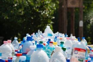 BPA inhibits adipocytes from releasing adiponectin, an adipokine that is a key regulator of insulin sensitivity[25] and helps to protect humans from obesity, diabetes, and heart disease.[26] BPA has been shown to affect the function of both the insulin-releasing β-cells and the glucagon releasing α-cells in the pancreas.[27] Animal studies have shown that exposure to BPA in utero may contribute to the development of diabetes in the offspring and aggravates insulin resistance associated with pregnancy in the mother.[28] In data analyzing 4,389 adults with diabetes, higher urinary BPA levels were associated with higher hemoglobin A1c (HbA1c) levels.[29] In another recent study, increased exposure to BPA was associated with insulin resistance in overweight or obese children.[30]