Researchers from the group of Hans Clevers identified and characterized rare cell types in the upper gastrointestinal tract. The researchers analyzed human biopsies from the healthy esophagus, three different gastric regions, and the duodenum, by single-cell analysis. The results, published in Cell Reports, identified rare cell types and provided insights into the cellular composition and gene expression patterns along the UGI epithelium under normal homeostasis. This cell type provides a link to gastrointestinal defects in patients with cystic fibrosis and identifies a rare cell type that is most likely responsible for the secretion of high volumes of water in humans.
Upper gastrointestinal tract and cystic fibrosis
In the publication in Cell Reports, the researchers from the Clevers’ lab describe how they used scRNA-seq to investigate the gene expression profiles of different cell types along the epithelium of the esophagus, stomach and upper part of the small intestine. To this end, they obtained biopsies from healthy human tissue and compared the obtained data to mouse datasets. Although the main aim of the paper was to characterize and describe the different cell types in these organs, the researchers also obtained several interesting findings. See IMAGES A,B,C below.
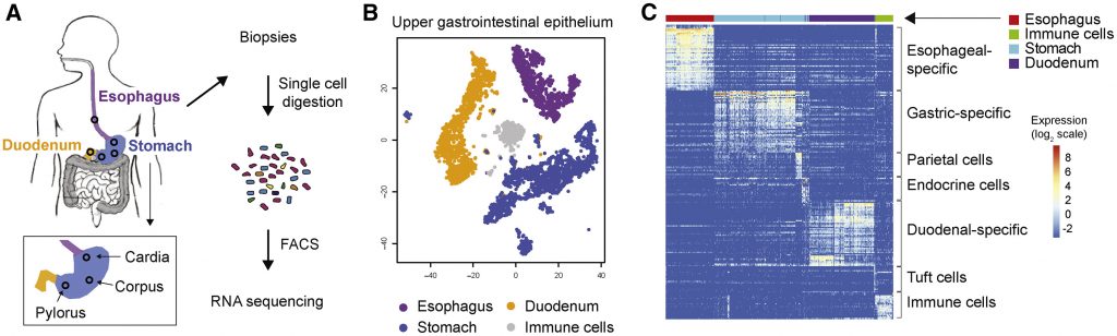
(B) t-SNE map of all combined scRNA-seq data displaying separate cell clusters for the esophagus (purple), stomach (blue), duodenum (orange), and immune cells (gray).
(C) Heatmap displaying the expression of the most differentially expressed genes, clustered according to their expression in the three UGI organs and parietal, endocrine, tuft, and immune cells.
Hormone-producing cells and histamine
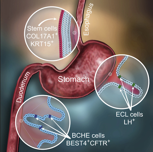
They found an esophageal stem cell population with high expression of the COL17A1 gene. Although mutations in this gene are known to lead to the development of blistering disease in the skin, defects associated with the esophagus are hardly reported in these patients. Additionally, the researchers characterized the cell composition in the stomach and found that the cells responsible for histamine production also express the Luteinizing Hormone (LH). This hormone is best known for its role in ovulation during the female reproductive cycle and for regulating testosterone levels in men. Why these cells in the stomach produce LH is unknown and requires further research.
Rare cell type in cystic fibrosis that leads to gastrointestinal obstruction
When studying the upper part of the small intestine, the researchers found a rare cell type with high expression of 4 genes, which are all linked to the secretion of high volumes of water. The most prominent member is the CFTR gene. Mutations in CFTR cause cystic fibrosis (CF). However, patients with CF also experience problems associated with gastrointestinal obstruction caused by a sticky, underhydrated mucus layer, which is most likely associated with the expression of the CFTR gene in the small intestine.
Translating results
Comparing human and mouse datasets for the gastrointestinal organs revealed differences in gene expression patterns between humans and mice. For example, the cells in the stomach producing LH were only seen in humans and the cell type secreting high volumes of water does not exist in mice. Overall, these differences were more extensive than initially expected. That complicates the translation of results obtained in mice to humans, and other methods need to be used to validate human-specific findings. “We could for example use organoids to follow-up on our current findings,” says Georg Busslinger, first author on the paper.
Conclusion/ Although the results described in the paper thus need functional confirmation, they bring the field a step closer towards knowing the exact cell composition in the gastrointestinal tract and what these cell types might do. “The results provide insight into the molecular characteristics of individual cell types and how they may function in the healthy epithelium. Moreover, these data serve as a resource for scientists everywhere,” Busslinger concludes.
Click Here for Full-Text Study